A new paper from the Taylor group at the University of Arizona introduces a metal-free, visible-light photosensitization strategy that labels Tryptophan residues with high selectivity in proteins, antibodies, and live cells – all run in the HepatoChem PhotoRedOx Box.
Tryptophan (Trp) is one of the least abundant amino acids in human proteins, appearing on average once per hundred residues. That scarcity, combined with its unique indole side chain, makes it an attractive handle for site-selective bioconjugation: labeling at Trp tends to be more site-specific than reactions at more abundant residues such as lysine or cysteine. Yet Trp-selective methods that work under fully aqueous, biologically compatible conditions and without metal catalysts have remained relatively limited.
A new paper in the Journal of the American Chemical Society from Vishal Agarwal, Hieu Pham, Samuel G. Bartko, and Michael T. Taylor at the University of Arizona and the University of Wyoming addresses this directly. Their work, “Cation–Cation Photosensitization for Protein Ligation and Intracellular Catalysis,” describes a purely organic photosensitization system that operates at 427 nm in aqueous buffer, requires no metal catalyst, and achieves protein labeling in minutes at micromolar reagent concentrations. The system performs across a range of substrates – from model proteins to therapeutic antibodies to live human cells – and all photochemical reactions were run in the HepatoChem PhotoRedOx Box.
The observation: one pyridinium activating another
The starting point was an unexpected finding. The Taylor group had been working on N-substituted pyridinium salts as reagents for Trp-selective ligation via photoinduced electron transfer (PET). During these studies, they noticed that pyridinium compound 1, when irradiated selectively at 427 nm in the presence of a second pyridinium salt (compound 2) that does not absorb at that wavelength, could nonetheless sensitize the reaction of compound 2. An excited cationic chromophore was activating a second cation that would otherwise be inert under those irradiation conditions.
The proposed mechanism begins with reductive quenching of the excited sensitizer by an exogenous electron donor (glutathione, GSH) to generate the reduced radical species 1•. This species then undergoes rapid single-electron transfer (SET) to pyridinium 2, generating the radical anion 2•. Pyridinium radicals of this class are known to fragment with exceptional speed, producing a short-lived carbamoyl radical that reacts with aromatic nucleophiles – primarily Trp residues in protein substrates – to yield carbamylated products.
The LUMO energy of the pyridinium acceptor turned out to be the critical controlling variable. When the group substituted compound 20 (the optimal pyridinium reagent) with a trimethylpyridinium salt (compound 21) whose calculated LUMO energy was 581 mV higher, labeling was completely abolished. The SET step is thermodynamically gated: only pyridinium salts with appropriately matched LUMO energies can accept an electron from the quinolinium radical anion efficiently.
Designing the optimal quinolinium sensitizer
The initial observation used the pyridinium compound 1 as both the sensitizer and the reactive species. To develop a genuinely catalytic system, the group sought a separate sensitizer scaffold that could drive the activation of stoichiometric pyridinium reagents at low loading. They identified 2,4-diaryl-N-methylquinolinium salts as a promising framework, on the basis of their tunable photophysics and demonstrated biocompatibility in prior work from the Chenoweth and Petersson laboratories.
Fifteen quinolinium sensitizers were synthesized and screened against three different pyridinium reagents across a range of sensitizer concentrations using lysozyme as a model protein. Clear structure-activity relationships (SAR) emerged. Sensitizers with strong dialkylamino electron-donating groups failed entirely. Sensitizers with moderately electron-donating methoxy (MeO) groups gave the highest labeling activity. Heteroaromatic substituents at the 2- and 4-positions gave little or no labeling. The optimal sensitizer, compound 16, bears methoxy-substituted aryl groups and outperformed all other structures tested, giving measurable labeling at concentrations as low as 5 μM.
Sensitizer 16 has photophysical properties well suited to this role: absorption extending to approximately 450 nm (λmax = 379 nm), a very large Stokes shift of 164 nm, a very low fluorescence quantum yield (below 0.01), and a photooxidation potential ([16+]*/16• of +1.4 eV). The low quantum yield is consistent with rapid non-radiative decay through the PET pathway rather than fluorescence, and the excited-state potential positions 16 well for reductive quenching by GSH and subsequent SET to the pyridinium acceptor.
From model protein to peptide to antibody
With the optimized 16/20 pair established, the Taylor group demonstrated labeling across a range of increasingly complex substrates, all using the HepatoChem PhotoRedOx Box.
Lysozyme served as the initial model protein. Irradiation of 10 μM lysozyme with 10 μM sensitizer 16 and 100 μM pyridinium probe 20 in pH 7.5 phosphate buffer for five minutes gave robust labeling, confirmed by intact protein mass spectrometry.
Leuprorelin, a therapeutic GnRH agonist peptide containing one Trp residue, was labeled at 91% conversion using only 10 mol% sensitizer 16 (10 μM) and 4 equivalents of pyridinium 20 (400 μM) in ammonium acetate buffer with 40 minutes of 427 nm irradiation. MS/MS of the labeled product confirmed modification at Trp. This result demonstrates catalytic turnover of the sensitizer at meaningful substrate concentrations.
Trastuzumab, the clinical anti-HER2 antibody, was labeled using azide-functionalized pyridinium reagent 20a, allowing installation of a clickable azide handle. In-gel fluorescence after Alexa Fluor 568 conjugation revealed modification primarily on the antibody heavy chains, and peptide mapping identified W99 on the heavy chain Fab region as the primary modification site. This level of site selectivity, combined with the mild conditions and aqueous buffer compatibility, is directly relevant to antibody-drug conjugate (ADC) development workflows.
Into the proteome: 319 proteins at 93% Trp selectivity
The group next moved to chemical proteomics in HEK293T cell lysate. Lysates were treated with sensitizer 16 (30 μM) and biotin-tagged pyridinium probe 20b (100 μM), irradiated for 20 minutes at 427 nm, and processed for streptavidin enrichment followed by LC-MS/MS proteomics.
The result was enrichment of 319 proteins with 93% selectivity to Tryptophan residues. The high Trp selectivity in a complex proteome is a meaningful demonstration of the chemoselectivity of the carbamoyl radical intermediate: even in the presence of all other nucleophilic amino acids, the reaction preferentially modifies Trp. Measurable labeling signal was detectable within one to five minutes of irradiation, consistent with the rapid kinetics observed in the isolated protein studies.
Live cell labeling: into the nucleus
The final demonstration moved into living HeLa cells. Cells were incubated with sensitizer 16 and azide-pyridinium 20a for 60 minutes to allow cellular uptake, then irradiated with 427 nm light for 20 minutes. Labeled cells were fixed, permeabilized, and reacted with dibenzocyclooctyne-conjugated Alexa Fluor 568 via strain-promoted azide-alkyne cycloaddition (SPAAC) for click-chemistry-based fluorescent imaging.
Confocal microscopy revealed labeling in multiple subcellular compartments, including nuclear regions. This is noteworthy because aromatic cationic compounds are generally assumed to accumulate in mitochondria on the basis of membrane potential-driven uptake. The observation of nuclear labeling suggests that the localization of these specific cationic structures is more nuanced and structure-dependent than simple lipophilic cation rules would predict. Live cell proteomics identified 101 enriched proteins, with gene ontology analysis pointing to enrichment of nuclear and nucleolar proteins.
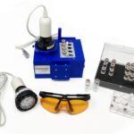
The HepatoChem PhotoRedOx Box as the photoreactor platform
All photochemical steps in this study – from the initial sensitization screen through the live cell labeling experiments – were performed in the HepatoChem PhotoRedOx Box (model HCK1006-01-016). The PhotoRedOx Box’s small vial format (2 mL Pyrex LC/MS vials) is well matched to the microliter-scale reactions used in protein labeling: reaction volumes are typically 50–500 μL, sensitizer and probe concentrations are in the low micromolar range, and reproducible, uniform illumination is essential for obtaining consistent mass spectrometry data across screening campaigns. The ability to run parallel reaction conditions within the same standardized photoreactor directly supported the SAR campaign across 15 sensitizer structures and three pyridinium probes.
Implications for protein bioconjugation and chemical biology
The Taylor group’s cation-cation photosensitization approach adds a genuinely new entry to the toolkit for site-selective protein modification. Several features distinguish it from existing approaches. It is entirely metal-free, removing concerns around metal contamination in biological samples. It operates at 427 nm under aqueous conditions at room temperature, making it compatible with sensitive biological substrates. It achieves 93% Trp selectivity in a complex proteome, a level of chemoselectivity that most amino acid-reactive chemistry cannot match in unfractionated lysate. The azide-functionalized pyridinium probe connects directly to established click chemistry workflows. And the demonstration in live cells, with imaging of the labeled compartment, opens the door to spatial proteomics applications where the photoreactor defines the labeled microenvironment.
For groups working on antibody functionalization, proximity labeling, or intracellular chemical proteomics, the 16/20 system described here represents a practical new option that merits evaluation alongside enzymatic labeling tools (APEX, BioID) and existing photoredox proximity labeling approaches.
Explore the HepatoChem PhotoRedOx Box
The photoreactor behind this chemistry. Standardized vial format, uniform illumination, compatible with any stir plate and Kessil LED sources.
Reference: V. Agarwal, H. Pham, S. G. Bartko, M. T. Taylor, J. Am. Chem. Soc. 2026, 148, 6750–6757. DOI: 10.1021/jacs.5c19565
Equipment used: HepatoChem PhotoRedOx Box (model HCK1006-01-016) with 427 nm Kessil PR160L LED.